a few experiments concerning vision

I wanted to see how perception happen as an operation of the nervous system. I was studying color vision, and I wanted to show how colors characterized in terms of spectral energies were coded in the retina of the person that recognized them. I had set up my experiments in those terms because in those times it was thought: “living systems received information about the colors in the medium through their eyes and coded it in the activity of the retinal neurons, to be decoded afterwards in the central nervous system.” I made many experiments and learned much about the activity of nerve cells, however I never could show what I thought I should be able to show, namely, that the activity of the retina correlated with the spectral composition of the colors projected on a screen. I spent three years doing this. Eventually I thought that maybe the question I was asking was wrong. Maybe the activity of retina did not correlate with the colors in terms of their spectral composition, but rather it correlated with the name of the color. My colleagues thought that I had become crazy.
But really, it was not such an insane idea. We know that we have illusions now and then. And when do we know that we had an illusion? In daily life we say that an experience that we live as valid in the moment that we live it was an illusion when we invalidate it afterwards as we compare it with another experience of which we choose not doubt.
… We speak as if a name were something abstract, external to us, and do not realize that the name refers to our feelings in the experience that we are living. Calling a person “John” or a color “blue” refers to what is happening in oneself, not to an entity supposed to be out there. So, when I name something I am referring to what is happening to me, to an internal dynamics of my nervous system. Thus when I proposed to correlate the activity of the retina with the name “of the color,” I was proposing to correlate the activity of retina with the activity of the rest of the nervous system that generated the act of naming. In doing this I began a new series of experiments to show that the activity of retina correlated with the name of the color. I was able to show this using a Skinner box and recording the activity of the retina using miniature electrodes.
A friend trained a pigeon to peck at different spectral colors for which we had standard names such as green, blue red … The pigeon would, for example peck at whatever we called “green”, regardless of whether it was spectral green, or a “color shadow” that we called green, even though its spectral composition was that of white light. This kind of experiment showed that the activity of the retina in chromatic perceptual experiences correlated with the name of the colored object, not with its spectral composition. And at the same time it showed that the nervous system operated performing internal correlations in which the name corresponded to a configuration of activity in the central nervous system.
Maturana 2012, Fundamentally Different
in press Constructivist Foundations
what the frog’s eye tells the frog’s brain
While Maturana was working on his PhD in Harvard in the 1960’s he participated in setting up some experiments which he later continued as a postdoc in MIT. What they were doing was recording the signals from individual ganglion cells in the frog’s retina and finding what kind of visual stimuli activity in these cells corresponded with. The resultant paper “What the Frog’s Eye Tells the Frogs Brain” is a classic, and it is how I first encountered Maturana’s work when I was still an undergraduate zoology student also in the 60’s.
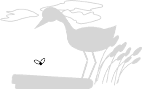
Both diagrams are selections from paintings by George P. Kelvin from Animal Behaviour,
Time Life Nature Library, 1965. The paintings and text were based on interviews with
H. Maturana and S. Frenk, University of Chile.
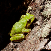
adequate for living
An amphibian such as a frog has a simpler nervous system than that of mammals. This made it easier to study, and it revealed clearly how much processing takes place as part of the visual experience. A frog has to deal with relatively simple things, like seeing where its going, avoiding a sudden movement of a large object such as a bill heading towards it, and catching food. Its visual system deals with all of these circumstances, much of it already in the retina itself.
frog retina
In this orientation, the light enters from below, and the top of the painting is where the pigment layer, the tapetum is located. I’ve arranged it in this orientation so I can tell the story in sequence.
-
1.The light sensitive cells are densely packed so that individual cells are stimulated or not according to the amount of light that impinges on them. Each cell branches and makes contact with
-
2.several bipolar cells; bipolar because they branch at both ends of the cell body. These in turn connect to a network of the third layer of cells, the ganglion cells in layer 3. Note that the connections between the sensors, the bipolar cells, and the ganglion cells have created a network wherein the specific architecture of connections propagates the original signal quite differently.
-
3.the ganglion cells, where Maturana and his colleagues measured the electrical activity are larger. Each has many branches (dendrites) that connect to the network of bipolar nerve branches, and then send a single very long axon off to the
-
4.optical nerve which leads to the brain, where the appropriate motor response is generated in what is known as a “reflex center.”
1
2
3
4
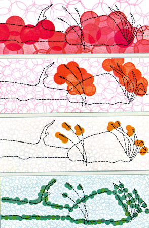
“Red ganglion cells, largest an least numerous of the four kinds, are affected by dimming: they respond only to darker parts of the swamp scene, such as shadows...
Orange ganglion cells, called “event detectors” become activated when movement [including the beak of a heron] occurs in the frog’s visual field...
Tan ganglion cells are set off by very small, moving objects with convex edges [such as the insects that frogs catch with a zap of their tongue] hence their name: the “bug detectors.”
Green ganglion cells, the so-called “edge detectors” react to sharp edges, either lighter or darker than the background.” [Together with the dimming receptors which indicate solid areas, they are useful to the frog for navigating its world]
Niko Tinbergen and The Eidtors of Life, in Animal Behaviour,
Time Life Nature Library, 1965.
my point:
The connections in the retina go a long way towards enabling the frog to adequately conduct its life in its normal medium.
The retina composes a “scene” of relevance for the frog.
the results
blue snow
Snow is white, not blue. Any spectrometer will tell us that. However, artists know something about human visual experience. They paint shadows in show using blue pigments, and it looks right to us. Similarly shadows on faces may be painted in greenish shades, and we don’t have green skin, and the green shadows don’t make us look sick.
What’s going on? Of course, it is all part of how our nervous system composes our experience; and how Maturana came to this understanding, of what he called “relativistic colour coding” is told in the following:
Jane Wolsak
Veronica Cordova de la Rosa
Paul Burman, KUMU Estonia